AngioScan-01 - Full description
“AngioScan-01” is a professional diagnostic device for assessment of arterial function state.
The developed method is based on monitoring of state of arterial function (stiffness of artery wall and state of endothelial function). Optical sensors operating in the near infrared spectrum and reliably recording pulse wave are used for these purposes.
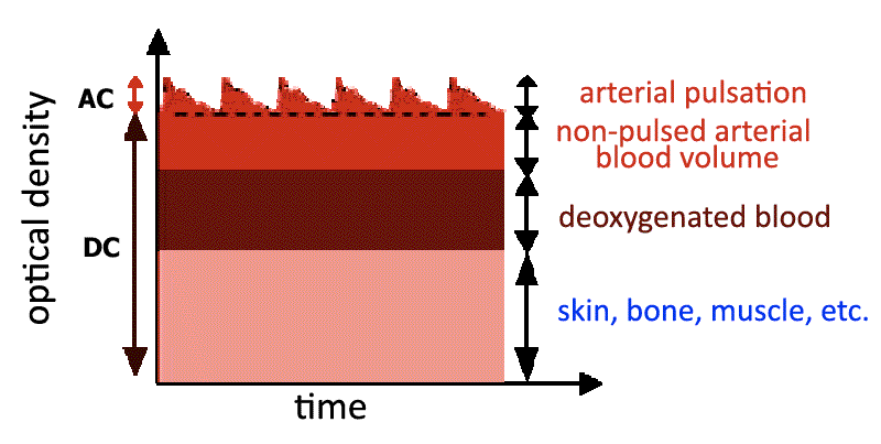
Hardware component of the device can be presented in a form of a specialized dual-channel photoplethysmography. Herewith we mainly focused on obtaining high-quality source signal while developing the device "Angioskan-01". Through the use of optical sensors with high dynamic range now we can register a permanent component of the signal (DC), associated with a constant optical density of tissue, as well as a variable component of the signal (AC), mainly determined by the activity of the heart (Pic. 1). The bandwidth of the photoplethysmography channel is from 0 to 30Hz. The frequency of digitization of the signal is 1000 Hz, which is determined by the need to measure time intervals with an accuracy of 1 ms. It is especially important when registering (the rate of retardation) a speed delay (phase shift) in the passage of the pulse wave while performing the occlusive test.

Pic. 1. Photoplethysmography signal formation diagram
It should be noted that in reality a scope of the variable component of the signal (АС) is limited to only a few percent of the total optical density.
Picture 2 schematically shows an optical sensor mounted on the terminal phalanx of the finger.

Pic. 2. Schematic representation of an optical sensor mounted on the terminal phalanx of the finger.
Infrared rays passing through the finger are recorded by a photo detector, which converts the light into a voltage (light/voltage converter) or frequency (light/frequency converter).
The signal is recorded either during the passage of photons from the light source to the photo detector through the tissue, or during the reflection, the light is reflected from the tissue back in direction of the photo detector. In the first case, the sensor is installed on the terminal phalanx of the finger, or earlobes, in the second it is installed with an adhesive layer on any part of the skin surface. Sensor operating in mode of light transmission has a better signal / noise ratio and it is more often used in pulse oximetry. Reflective sensor has two main advantages: no restriction on site and minimum compression of the tissue segment. The reflective sensor has significant advantages in cases when the signal is to be monitored during a long period of time (while intensive therapy or observation of a patient), as it is necessary to change the location of sensor, made in a form of “peg”, only after a several hours of test. However, the sensors operating in the mode of light transmission are optimal in case of short-term (few minutes) testing.
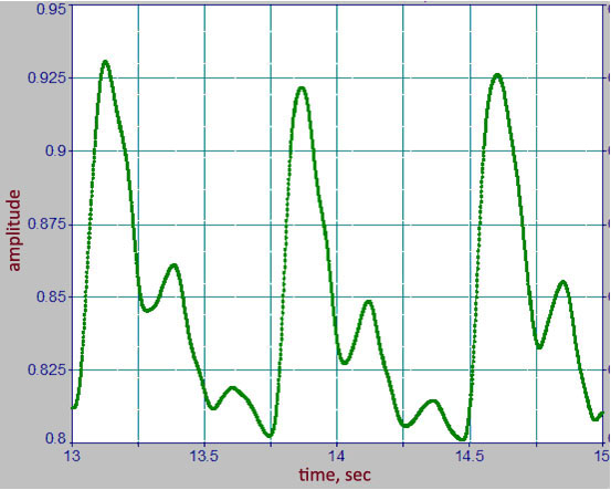
Picture 3 shows pulse waves recorded by the optical sensor mounted on the finger.

Pic. 3. Example of typical volume pulse waves.
Assessment of the state of endothelium
Functional (occlusal) and pharmacological tests are used for assessment of the state of endothelial function. While developing the devices we tried to eliminate influence of an operator on test results and to provide an ease of use, so now the devices can be easily used in medical centers, as well as at home.
The registration technology we developed and contour analysis of volume pulse wave make possible to obtain clinically important data about the stiffness of the elastic type arteries (the aorta and its main trunks). Original photoplethysmogram signal processing algorithms assess the following parameters:
- Duration of the expulsion of blood by the left ventricle;
- Amplitude and timing of early and late systolic wave;
- Augmentation index (or effect of the reflected wave);
- Assessment of the health of baro-receptor sensor system;
- Value of central arterial blood pressure.
Occlusion test, carried out by using the cuff mounted on the shoulder, registers the state of endothelial function:
- In small resistive arteries (microcirculation system);
- In large conductive arteries.
It is possible to evaluate the vasomotor response to exogenous nitric oxide by conducting pharmacological test with nitroglycerin. Respiratory test can identify individuals with severe atherosclerosis of the coronary arteries.
Evaluation of stiffness of the arterial wall
The registration technology we developed and contour analysis of volume pulse wave make possible to obtain clinically important data about the stiffness of the elastic type arteries (the aorta and its main trunks). Original photoplethysmogram signal processing algorithms assess the following parameters:
- Duration of the expulsion of blood by the left ventricle;
- Amplitude and timing of early and late systolic wave;
- Augmentation index (or effect of the reflected wave);
- Assessment of the health of baro-receptor sensor system.
Assessment of central arterial blood pressure
In recent years it has been found that blood pressure (BP) in the aorta (central blood pressure) reflects the blood flow in the coronary and cerebral vessels, and it is a more reliable predictor of cardiovascular risks rather than blood pressure, traditionally measured at the shoulder. A measurement of blood pressure in the carotid artery can be used for assessing of the central pressure, since it corresponds to a pressure in ascending aorta. The recording of pulse wave in the carotid artery, performed by tonometry sensors, can be used as a direct testing (BP) method followed by measuring of a pulse pressure. The calibration of arterial pulse is based on state of the mean and diastolic (BP) tones, which show constant parameters in different vascular beds.
We have developed an algorithm for evaluation of the central arterial blood pressure. The central arterial blood pressure is assessed by a standard tonometer before the contour analysis.


